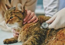
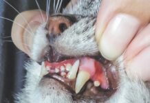
During your cats lifetime, any of a host of deeply seated physical ailments might threaten her life. Many of these disorders might be immediately recognizable to your veterinarian during the course of a routine, superficial examination. Others would be apt to remain a mystery, with diagnosis achievable only by means of an imaging technology that provides the practitioner with a detailed view of the animals bones, muscles and organs. Four such imaging modalities are now used in veterinary medicine – X-rays, ultrasound, computed tomography (CT) scanning and magnetic resonance imaging (MRI). Which among them will be most suitable for your cat, says Peter Scrivani, DVM, an assistant professor of veterinary imaging at Cornell Universitys College of Veterinary Medicine, depends primarily on which tissues and organs in the animals body need to be examined.
The responsible cat owner can benefit from a basic understanding of these imaging methods – their underlying technology, their virtues and limitations, their availability and other relevant matters, such as the cost of a typical procedure. Following are brief accounts of the four imaging modalities, presented in order of the frequency with which they are currently used by veterinarians.
X-Ray Imaging
Relied upon by veterinarians for the past half-century, this technology – also called radiography – employs an invisible and highly penetrating light ray. Dr. Scrivani describes the procedure, the most commonly used of all veterinary imaging modalities, as follows:
We direct an X-ray beam through the patients body, focusing on the area that we want to examine. The rays pass through the body and onto a film. Different tissues absorb varying amounts of the radiation. If a tissue absorbs a lot of X-rays, fewer of them will reach the film, and that part of the image will be white or a light shade of gray. If no rays are absorbed, that part of the film will be black.
Due to their density, bones absorb most of the rays and therefore appear white on a radiographic image, while soft tissues – which absorb few or none of the rays – will be black or shades of gray. Thus, X-rays are an especially effective medium for viewing bones – studying fractures and monitoring their healing, for example. This technology is also useful for diagnosing such skeletal disorders as osteoarthritis, hip dysplasia and bone tumors. In addition, X-rays are often used to examine an animals lungs, since these organs are normally filled with air, which allows a directed beam to pass through to the film.
An X-ray examination, says Dr. Scrivani, will usually take no more than a half-hour, and the procedure is comparatively inexpensive – about $100 for a set of X-rays of, say, the thorax or abdomen. The appropriate equipment is available at the vast majority of veterinary clinics. And many patients do not require sedation.
The chief limitation of X-rays, Dr. Scrivani points out, is that the images are flattened together – they dont have a third plane. Due to the lack of contrast resolution, the ability to differentiate one tissue type from another may be inferior to what we get with other imaging methods.
Ultrasound
In ultrasonic imaging, a technology that has been used by veterinarians since the late 1970s, high-frequency sound waves, well beyond the range of hearing, are directed at a specific area of an animals anatomy. When the sound waves bounce back from the target area, they are transmitted to a central processing unit that instantaneously assembles this information to create a cross-sectional visual image – a sonogram – that appears on a display monitor.
Ultrasound is superior to X-rays for viewing soft tissues and organs, such as the liver or bladder, as it can differentiate tissues and their associated fluids.

324
Ultrasound is the one modality that gives us cross-sectional images without requiring the patient to be under general anesthesia, says Dr. Scrivani, although some animals may have to be somewhat sedated if theyre too aggressive or if theyre in pain. The procedure typically lasts about a half-hour he says, and is free of any hazards.
A sonogram often yields a wedge-shaped image, since that is the shape of the sensing mechanism at the tip of the probe that is frequently used to pick up sound waves during the procedure. To the untrained eye, a sonogram is likely to be nothing more than a mottled field of black, white and gray. To experienced radiologists, however, the image can provide a wealth of information about an animals vital organs. A form of ultrasound called echocardiography is frequently used to view the structure of an animals heart.
Ultrasound, the second most commonly used veterinary imaging modality, is currently available at large referral hospitals and specialized clinics, as well as at smaller veterinary clinics that have the space for the equipment and staff who are trained in its use. An ultrasound examination of the abdomen, says Dr. Scrivani, will cost about $200.
CT Scanning
The concept behind conventional X-ray technology also applies to CT scanning. That is, X-rays are beamed through target areas of the patients body, with different tissues absorbing varying amounts of the rays. But in CT, the patient lies on a movable table that slides slowly into the donut-shaped portal of a cylindrical, rapidly spinning X-ray projector. A series of exposures are made as the projector rotates around the patients body, sending narrowly focused X-rays through to detectors on the opposite side of the cylinder. The accumulated data are immediately transmitted to a computer, which processes the information and yields, almost instantaneously, a cross-sectional image of the area under study.
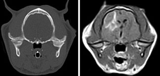
The chief advantage of CT over conventional X-rays, says Dr. Scrivani, lies in its ability to produce images that are cross-sectional and that differentiate more types of tissues. This advantage is due to the fact that the contrast resolution in CT scans can be manipulated by a technician during an examination.
CT is well suited for producing precise images of organs lying within an animals thoracic cavity. This imaging modality is often used in cases of compressive spinal cord disorders, abnormal tissue growths, and conditions affecting an animals skull, brain, ears or nose.
To avoid a blurred image, patients undergoing a CT scan must remain motionless during the procedure and therefore must be under general anesthesia. The duration of the procedure varies. The imaging itself takes just a few minutes, although the entire procedure may take a half-hour or so due to the careful set-up time that is required. The cost of a CT scan, says Dr. Scrivani, will typically be about $500.
The availability of CT scan technology is currently limited to large veterinary referral hospitals and specialty practices, he notes.
MRI
No veterinary imaging modality is more complex than MRI, which has been used by veterinarians since the mid-1990s. For this type of examination, the patient is placed on a table centered within a very strong magnetic field, which causes subatomic particles (protons) in the patients body to line up and spin in a particular manner. When the protons are so arranged, technicians shoot a radio wave into the patients body, which deflects the spinning particles. Then, when the radio wave is shut off, the protons return to their natural state and, in doing so, release energy. This released energy creates signals that are picked up by an antenna and fed into a computer, which interprets the signals and produces precise cross-sectional images of the patients internal organs and tissues in clearly distinguishable shades of black, white and gray that can be viewed on a computer monitor.
According to Dr. Scrivani, MRI provides better cross-sectional resolution than other modalities and is thus more accurate in diagnosing certain disorders, such as tumors, spinal cord diseases, neurologic conditions and disorders associated with soft tissues in the joints.
There are two drawbacks involved with MRI: (1) animals must be under general anesthesia for the procedure, which usually takes 45 minutes to an hour; and (2) the procedure is comparatively expensive. Typically, says Dr. Scrivani, an MRI exam will cost about $1,000. He points out as well that MRI imaging is currently offered only at large veterinary referral hospitals or at private facilities that specialize in the technology.


