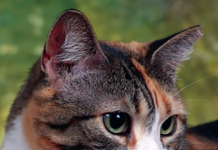
Cats eyes are spherical in shape and rest in bony sockets in the skull, cushioned by fat pads. Muscles connect the eye to the socket, allowing movement in various directions. Upper and lower eyelids, plus a third eyelid, close to protect the eye. Lubrication is provided by two lacrimal (tear) glands, one tucked under the upper eyelid and one at the base of the third eyelid. There is confusion in language when people say tear ducts, says Matthew Chavkin, DVM, of the Veterinary Referral Center of Colorado. Lacrimal glands secrete tears that travel through numerous, microscopic ducts, bathe the eye, and then drain into the nasolacrimal duct which empties into the nose or the roof of the mouth.
What can a cat see?
Cats vision is somewhat fuzzy; they are unable to see fine detail and are unable to focus within six to eight inches of their eyes. Compared to humans, cats dont have good vision. Theyd be legally blind by our standards, says Chavkin. On the other hand, cats can see much better in dim light than we can.
Looking directly into your cats eyes, you see the cornea – the crystal-clear front covering of the eyeball, which is surrounded by white called the sclera. Over the sclera is the conjunctiva that also covers the inner surface of the eyelids and both sides of the nictitating membrane (third eyelid) but not the cornea. Behind the cornea is a chamber filled with watery liquid, aqueous humor; the pupil, surrounded by the iris; and the lens, which is held in place by muscles and ligaments. Like the cornea, the lens should be clear, says Chavkin, who is a 1990 graduate of the Cornell University College of Veterinary Medicine and is board certified by the American College of Veterinary Ophthalmologists. But every cat over the age of 8 develops lenticular sclerosis, a normal age-related hardening of the lens. In older cats, the pupil looks hazy, and people often think the cat has trouble seeing or has cataracts, which may not be the case. Located behind the lens is the vitreous humor, a jelly-like liquid that fills 70 to 80 percent of the eye and is largely responsible for maintaining eye shape.
The backside of the interior eye is the retina with light-sensitive rods (black and white/light and dark sensors) and cones (color sensors). Cats have color vision, but cannot differentiate reds from green very well. Their retinas are primarily rod retinas – rods are more sensitive and are designed for nocturnal vision. Behind the retina is the reflective tapetum lucidum that reflects light not absorbed on its first pass through the retina. This helps with night vision. The optic nerve exits the back of the eye and transmits signals to the brain.
Nocturnal vision
Although there are more similarities than differences between the human and the cat eye, says Chavkin, one difference you notice right away is the third eyelid which rests on the eyeball at the inside corner. This thin structure is protective and helps lubricate and cleanse the eye. When visible, its often an indication there is something wrong with the eye.
Another difference from humans is in the cornea, which is about twice the size proportionally of the human cornea, and the pupil is a vertical slit rather than round. “This is common among nocturnal animals, adds Chavkin. The cats retina, the light sensitive membrane at the back of the eye, contains many more rods than cones (about 25 to 1 in cats, 4 to 1 in humans), assisting dim light vision. Finally, the human eye lacks the tapetum lucidum, which creates the eye shine seen in cats eyes – more visible at night because the pupils are wide open. This specialized layer is present in most nocturnal animals and in all cats.
Contrary to the popular myth, cats cannot see in total darkness. There must be some small amount of light for the cat to see. It may appear totally dark to us, but it would not be for the cat. Cats have better vision in low light than people do, but in regular room light or in daylight, our visual acuity is six to eight times better than the cats, Chavkin says.


